What causes SCAD? This is one of the key questions SCAD researchers around the world are working hard to answer. Understanding the mechanism of how a SCAD occurs is an important aspect to advance knowledge and guide further research.
The European Position Paper published in February 2018 describes two potential mechanisms:
1) ‘Inside-out’: the development of a ‘tear’, allowing blood to accumulate in the artery;
2) ‘Outside-in’: the development of a ‘bruise’ that haemorrhages (leaks) into the artery wall.
To investigate these theories further, the UK research team led by Dr David Adlam collaborated with colleagues throughout Europe and in South America to study intracoronary images of SCAD patients obtained via Optical Coherence Tomography (OCT).
OCT is a kind of intravascular ultrasound that utilises light to deliver high-resolution cross-sectional images of the inside of the blood vessels. These images are more detailed than the images obtained during a coronary angiography, which is the more routine imaging method used to investigate the heart. Interventional cardiologists may perform OCT in addition to aid decision making for the optimum treatment plan for a patient.
To date, SCAD has largely been thought of in the context of the first mechanism, that a tear develops which blocks normal blood flow and often leads to a heart attack. However, the study found that a proportion of SCAD patients had no visible tear at all. The study also provides insight into the effects of pressure due to compression of the blood vessel which could mean that the tear develops as a result of the pressure rather than the tear being the primary event.
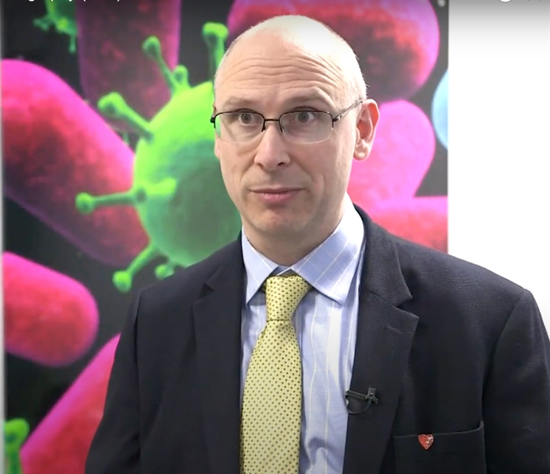
In this video, Dr Adlam describes the study, summarises the findings and their importance, in addition to describing next steps for the research.
The paper, Spontaneous Coronary Artery Dissection: Pathaphysiologal Insights from Optical Coherence Tomography, was published by JACC: Cardiovascular Imaging in March 2019.