Connective tissue abnormalities, coronary inflammation and coronary artery Fibromuscular Dysplasia (FMD) have all been suggested as potential causes of SCAD, but have not, until now, been systematically assessed.
This recently published paper, Vascular histopathology and connective tissue ultrastructure in spontaneous coronary artery dissection: pathophysiological and clinical implications is the largest study so far looking at SCAD coronary histopathology (the changes in tissues caused by disease). It is also the first systematic assessment of skin collagen ultrastructure, which is the fine structure that can only be seen at very high magnification, in SCAD patients.
Connective tissue
Although SCAD is associated with hereditary connective tissue disorders in a small number of cases and some SCAD patients display features of hypermobility, leading to theories that connective tissue abnormalities may be a cause of SCAD, the researchers didn’t find general differences between the skin connective tissue of SCAD patients and healthy volunteers. When looking at the elastin (a protein-forming the main constituent of elastic connective tissue, found especially in the dermis of the skin), the study found the suggestion of some subtle features of elastin damage might be different between healthy volunteers and SCAD survivors. The authors say these findings are helpful in identifying theories they can investigate further.
These findings suggest that any connective tissue abnormalities common to patients with SCAD who do not have hereditary connective tissue disorders are probably subtle, transient or localised to the coronary arteries.
Inflammation
The authors conclude that in terms of inflammation, it is likely that SCAD causes an inflammatory response in the coronary artery wall as the body tries to heal itself, rather than inflammation being a cause of SCAD.
FMD
An association between SCAD and FMD in non-coronary arterial beds is well established but it is less clear if the microscopic changes in the arrangements of cells in the arterial wall of patients with FMD are also found in the coronary arteries of patients with SCAD. This study did not find evidence that changes like those described in FMD commonly occur in the coronary arteries of SCAD patients suggesting these findings are not a universal finding in SCAD and may not therefore be the primary cause of a coronary arterial weakness predisposing to SCAD in most patients.
Outside-in or Inside-out?
The study concludes that the immediate cause of SCAD is likely to be the development of a spontaneous intramural haematoma (the ‘outside in’ hypothesis) rather than an intimal disruption or ‘tear’ (the ‘inside out’ theory). This does not seem to be directly related to increased density of the small blood vessels that supply the walls of large blood vessels (vasa vasorum), coronary FMD or local inflammation (except as a response to injury).
SCAD in autopsy
The authors say that in cases of Sudden Coronary Death (SCD), it is important to assess coronary arteries during a post-mortem to exclude SCAD, especially where there is no evidence of myocardial necrosis (irreversible death of heart muscle following a lack of oxygen). And in cases where there is no sign of a heart attack, careful assessment should be made of the distal arteries for evidence of SCAD.
Low rates of atherosclerotic changes were seen in the autopsy cases. This may be because the SCAD patient population is mainly young/middle-aged females whose risk of atherosclerotic disease is low, but is also consistent with recent findings suggesting a common genetic variant (PHACTR1/EDN1) seen in SCAD patients reduces the risk of ischaemic heart disease.
UK research
Thanks to everyone who contributed to this research, including those who have signed up for the UK research project, especially those who provided skin samples for analysis. Everyone who has allowed the researchers to access their medical records, study their scans and analysed their blood or skin samples is helping us get closer to answers, so please know that you are making a major contribution! If you haven’t yet signed up, please do.
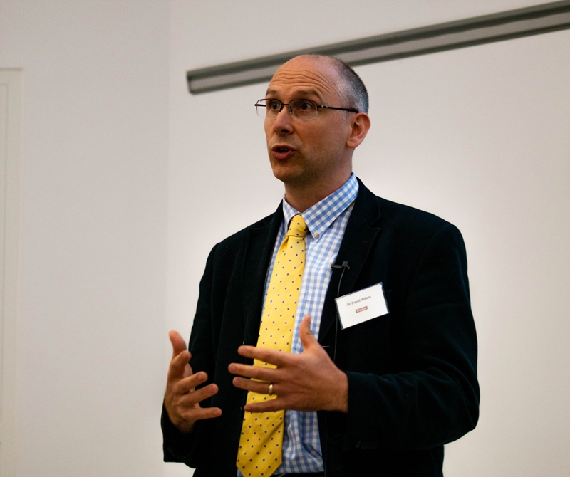
The paper was a collaboration between researchers working on the UK SCAD project, including Dr David Adlam, plus Dr Mary Sheppard from St George’s Medical School in London, Jan Lukas Robertus from the Royal Brompton, Sarah Parsons from Monash University, Melbourne Victoria, and Jan von der Thüsen from Erasmus MC, University Medical Center Rotterdam, Rotterdam, The Netherlands.
Beat SCAD helped fund this research.

